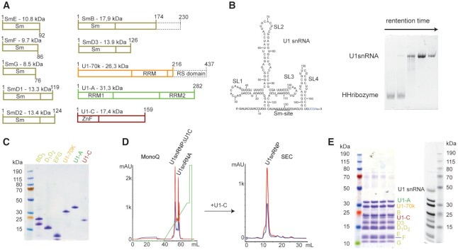
Figure 1.
Reconstitution of U1 snRNP. (A) Scheme of the U1 snRNP protein components. Dashes boxes correspond to the parts that were not included in our constructs. (B) Scheme of the U1 snRNA used for this study. On the right, a 10%-acrylamide urea PAGE stained with toluidine illustrates the separation between the hammerhead ribozyme and the U1 snRNA using purification in denaturing conditions. (C) SDS-PAGE stained with Coomassie showing the purified U1 snRNP protein components. (D) Purification of U1 snRNP. MonoQ was used as the anion exchange chromatography column and SEC stands for size exclusion chromatography. (E) SDS-PAGE gel stained with Coomassie (left) or with silver nitrate (right) showing the purified U1 snRNP.

