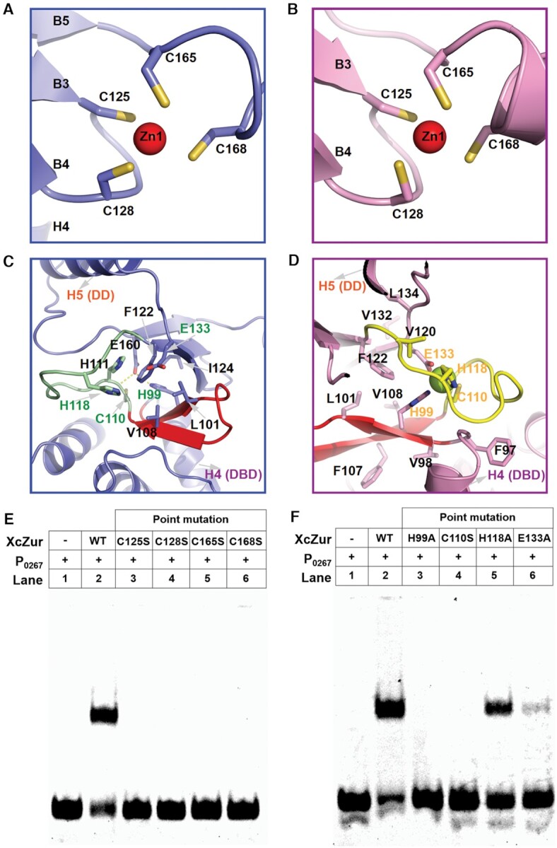
Figure 5.

The structural and regulatory zinc binding sites of XcZur. (A) Close-up view of the structural zinc binding site (site 1) of apo XcZur. The zinc atom is represented by a red sphere and the corresponding coordination ligands are shown in colored sticks. (B) Close-up view of the structural zinc binding site of holo XcZur. (C) Close-up view of the regulatory zinc binding site (site 2) of apo XcZur. Key residues in this site are shown in colored sticks. The characteristic β hairpin in DBD are shown in red. (D) Close-up view of the regulatory zinc binding site of holo XcZur. (E) Binding activity of wild type and site 1-related mutants of XcZur with P0267. (F) Binding activity of wild type and site 2-related mutants of XcZur with P0267.
