Figure 7.
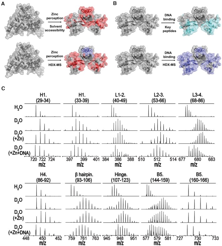
In solution evidence for zinc-mediated conformational changes of XcZur. (A) Peptides with significant changes in solvent accessibility (upper panel) or deuterium exchange (lower panel) upon zinc perception are mapped onto the crystal structures. (B) Peptides that contain residues important to DNA binding are mapped onto the holo-XcZur structure (upper panel). Peptides with remarkable changes in deuterium exchange during DNA binding are mapped onto the holo-XcZur structure (lower panel). For (A) and (B), the more exposed and hidden peptides are colored in red and blue, respectively. Key peptides for DNA recognition are colored in cyan. (C) Representative mass spectra for XcZur peptides from HDX-MS.

