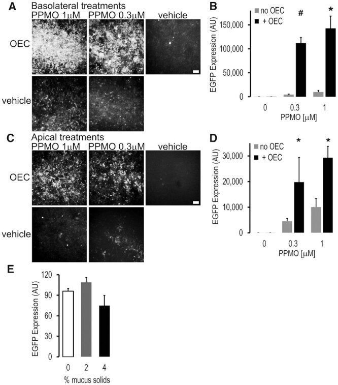
Figure 2.

Correction of a splicing mutation in mouse-derived primary airway cells using PPMO plus OEC. Primary mouse tracheal epithelial cells derived from the EGFP654 mouse (EGFP654 MTEC) were differentiated on permeable supports under air-liquid interface for 2–3 weeks as described in Methods. (A–D) PPMO plus OEC effects. The SSO PPMO654 and OEC7938 were administered once in a bolus either into the basolateral (A, B) or apical (C, D) compartments; PPMO (0.3 and 1 μM, 16 h) followed by OEC7938 (10 μM, 2 h). MTEC were subjected to microscopy and fluorometric analysis 48 h post-treatment. Live MTEC were imaged with constant settings in a Leica DMRB fluorescent microscope using a 10x magnification, bar = 100 μm (A, C), and quantitated in a Azure fluorimeter scanner (B, D). All wells have similar total cell numbers; mean ± SD; n = 3; *P < 0.01 and #P < 0.05 versus no OEC. (E) Effect of apical mucus on PPMO plus OEC delivery in EGFP654 MTEC. PPMO (0.5 μM) and OEC (10 μM) were administered apically mixed in a small (30ul) bolus of human mucus at 2 and 4% solids’ concentration; 0 = cell medium bolus; other experimental conditions were as before. Fluorescence was quantified in a Tecan system with a control set at 100%; mean ± SD, n = 4. Lumenal mucus did not greatly affect delivery of PPMO and OEC to the airway epithelial cells and thus splicing correction and EGFP expression was observed.
