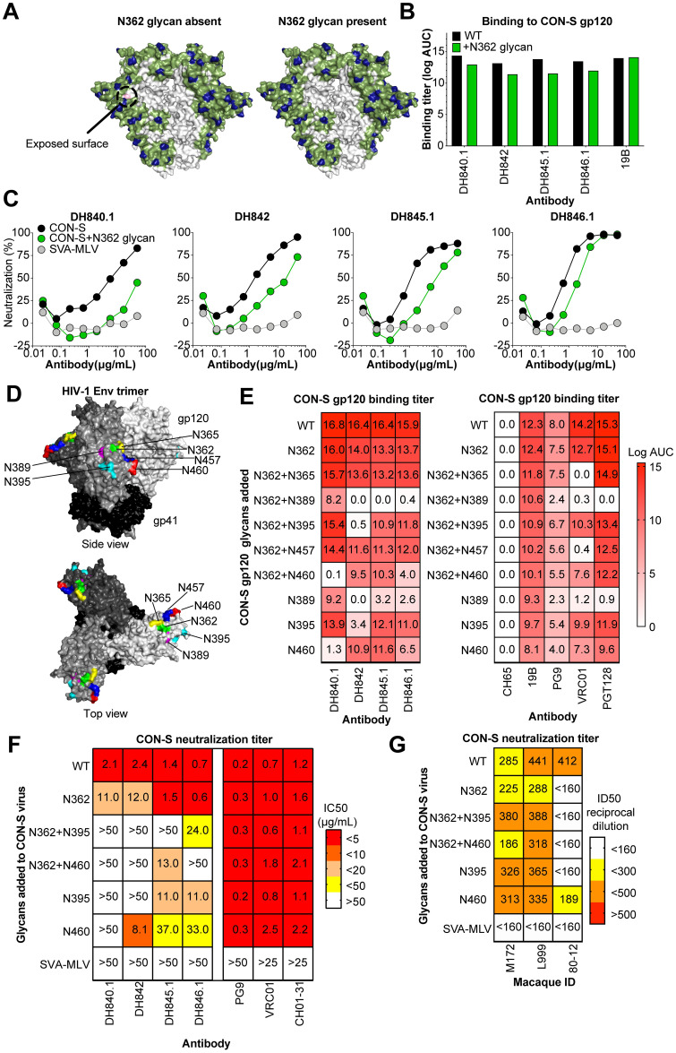
Fig 3. Hyperglycosylation of HIV-1 envelope determines vaccine-induced CON-S nAbs bind the distal side of the HIV-1 envelope gp120 subunit.
(A) Computational prediction of the CON-S envelope glycan coverage of HIV-1 envelope surface using the Glycan Shield Mapping tool on the Los Alamos HIV Database (https://www.hiv.lanl.gov/content/sequence/GLYSHIELDMAP/glyshieldmap.html). Two protomers of the trimeric envelope is shown in surface representation with potential N-linked glycosylation sites highlighted in blue. The surface potentially covered by a glycan attached to the glycosylation site is shown in green assuming 10 angstrom radius of coverage by each glycan. The gray surface in the center is receding inwards towards the trimer axis, and is predicted to be glycan unshielded; however, it may not be easily accessible to antibodies due to conformational masking from other protomers. (Left) The light pink indicate surface that is covered in 50–80% of group M HIV-1 isolates, but not covered in CON-S. (Right) The addition of a glycosylation site at N362 covers the exposed surface when occupied with glycan. (B) Vaccine-elicited neutralizing antibody binding to CON-S gp120 with or without the N362 glycosylation site. Binding titers are shown as log AUC as described in Fig 2. 19B, a V3 region-specific antibody, was used as a positive control. Mean values from two independent experiments are shown. (C) Monoclonal antibody neutralization of wildtype (black) and N362 glycan-modified (green) HIV-1 CON-S infection of TZM-bl cells. Murine leukemia virus was used a negative control. Representative results from 2 independent experiments. (D) The sites of novel glycan addition in C3, C4, V4, and V5 to block monoclonal antibody binding to CON-S gp120 are shown on the structure of trimeric HIV-1 envelope (PDB:5FYL). Each gp120 of the trimer is colored a different shade of gray and gp41 is colored black. (E) Antibody binding titers as log AUC are shown for CON-S gp120 wildtype and hyperglycosylated variants. The glycosylation site added is shown for each row. V1V2-glycan (PG9), CD4 binding site (VRC01), and V3-glycan (PGT128) bnAbs were examined for comparison to vaccine-induced macaque antibodies. 19B is a V3 region-specific antibody. The anti-influenza antibody CH65 was used as a negative control. Mean values of two independent measurements are shown. (F) Monoclonal antibody neutralization of infection of TZM-bl cells with wildtype CON-S and CON-S with specified Env glycan additions. Neutralization titers are shown as IC50 in μg/mL and color-coded based on the legend. (G) Plasma neutralization of infection of TZM-bl cells with CON-S pseudoviruses shown in (F). Neutralization titers are shown as ID50 reciprocal plasma dilution and color-coded based on the legend.

