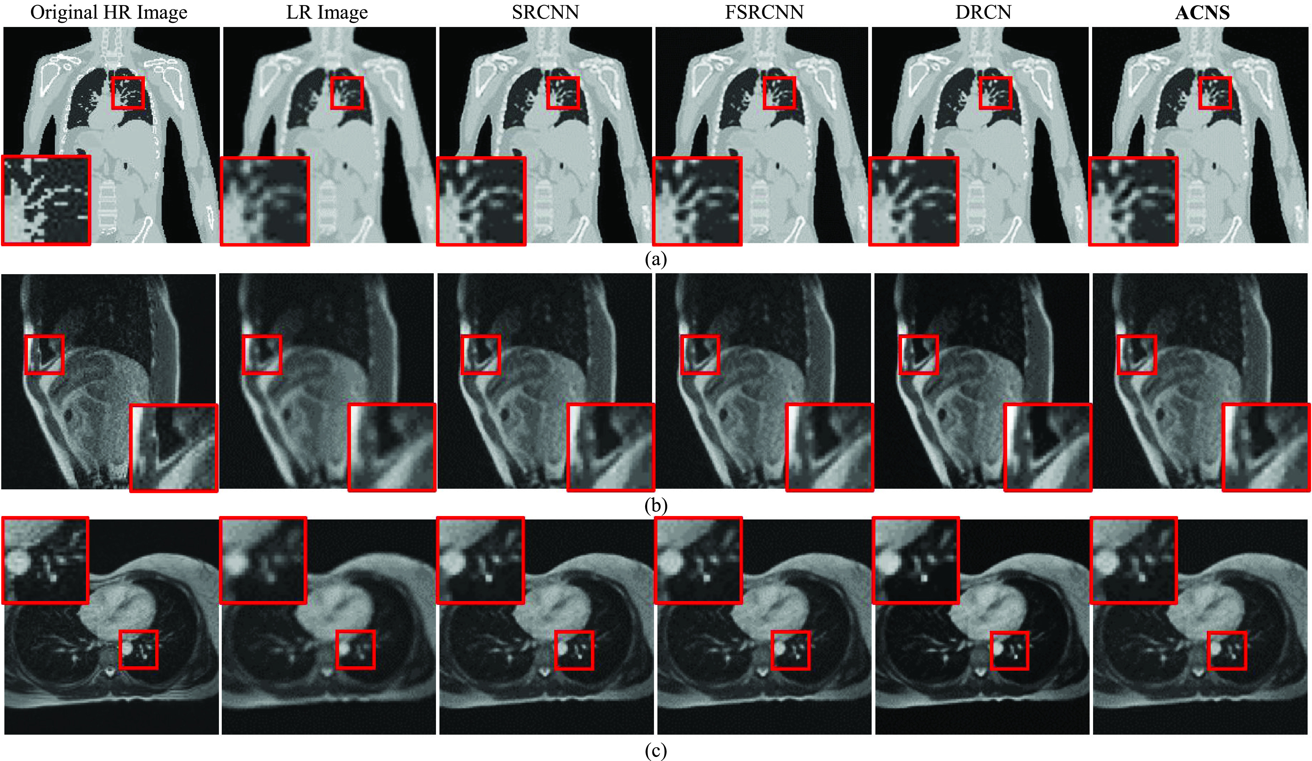
FIGURE 5.

Comparison of resolution-enhanced XCAT image and MRIs using SRCNN, FSRCNN, DRCN, and ACNS: (a) XCAT dataset1, (b) Volunteer1, and (c) Volunteer2 datasets. The first and second columns present the original HR and LR images; and the third, fourth, and fifth columns indicate SR results from SRCNN, FSRCNN, and DRCN, respectively. The sixth column is the resolution-enhanced XCAT image and MRIs by ACNS. All results were at a  of 2. To provide visible comparison, the images were partially enlarged from the regions marked as red squares.
