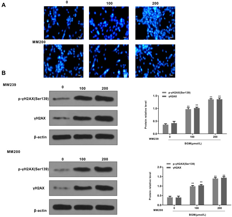
Figure 4.
BGM induces DNA damage in the melanoma cells. (A and B) The WM239 and MM200 cells were treated with BGM at the indicated dose. (A) The DNA condensation was analyzed by the 4, 6-Diamidino-2-phenylindole (DAPI) staining in the cells. (B) The γH2AX phosphorylation (Ser139), expression of γH2AX and β-actin were measured by Western blot analysis in the cells. The results of Western blot analysis were quantified by ImageJ software. Data are presented as mean ± SEM. Statistic significant differences were indicated: **P < 0.01.

