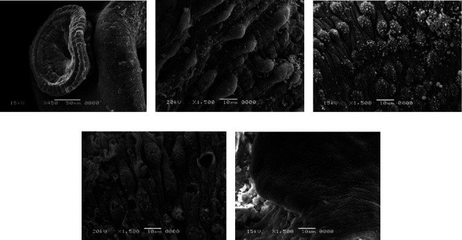
Figure 11.

Scanning electron microscopy of S. haematobium (adult worms and schistosomula) after exposure to different concentrations of Salvia fruticosa. (a) Destroyed sucker. (b, c, d) Dorsal surface of male showing tegumental exfoliation with damage and exfoliation of spines and tubercles. (e) Schistosomula.
