
Figure 2.
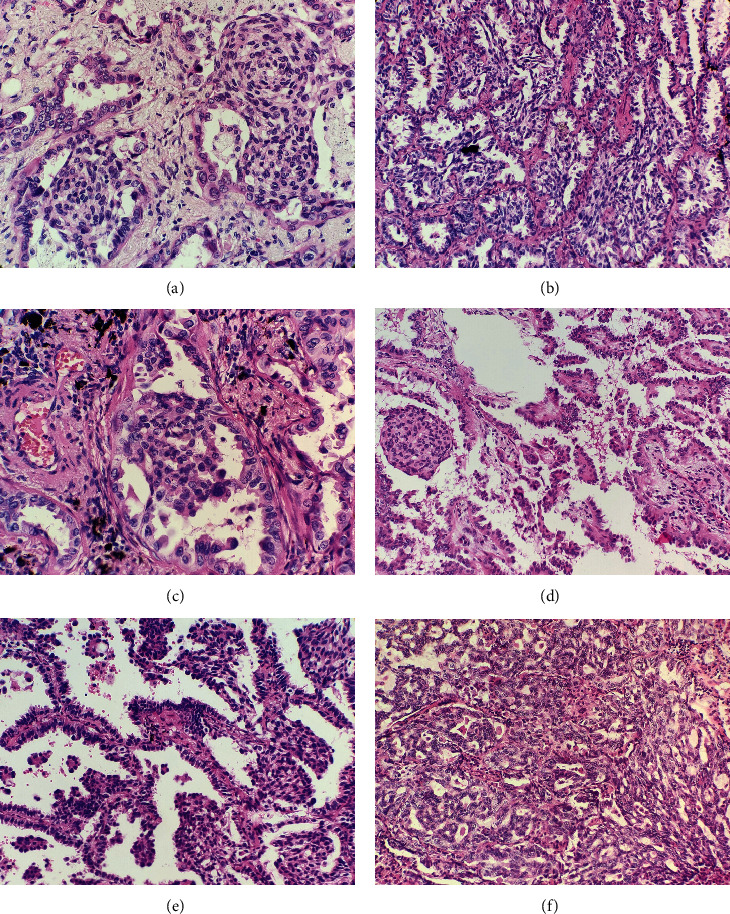
Histological findings of lung adenocarcinomas with MLCs. The MLCs were composed of spindle cells showing a whorled growth pattern, and the spindle cells in the MLCs had a syncytial appearance (a). The MLCs showed a streaming growth pattern with fenestration which was similar to the usual ductal hyperplasia in the breast (b). A few MLCs were epithelioid (c). An MLC in a papillary adenocarcinoma (d). MLCs and micropapillary components in a lepidic adenocarcinoma (e). The transitional region between the cribriform component and the MLC (f).