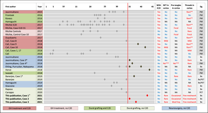
Fig. 2.
Visualisation of iatrogenic Aβ pathology incubation times in the current and in published studies. Left columns, first author and publication year, and case ID in the respective publication, where indicated. Centre, timeline of reported incubation times. Each diamond indicates a published case. Cases with incubation times of 35 and more years are highlighted in dark grey (published) and red (this study). The reported presence of tau pathology is indicated in the four columns on the right. The column on the far right indicates the sample type (Bx—diagnosed on biopsy; PM—diagnosed on post-mortem material; PET—diagnosed on in vivo PET imaging). * (leftmost column) indicate three comparison cases shown in Fig. 1; ** (column “threads in cortex”) highlight cases, where rare neocortical threads or granular tau pathology were reported in the context of abnormal prion protein pathology; *** (columns “NFT (neurofibrillary tangles) in cortex” and “pre-tangles in cortex”) corresponds to a case in which tau pathology is seen in the medial temporal lobe but not in the neocortex. °For case 1 it is unknown if a cadaver-derived dural graft was used during neurosurgery

