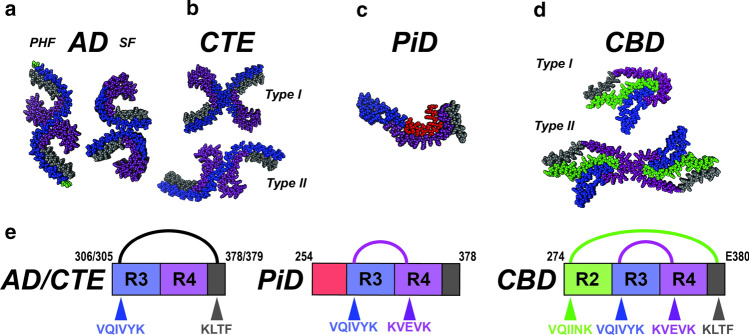
Fig. 5.
Unifying themes for diverse tauopathy fibrils. a–d Cryo-EM structures of tau fibrils isolated from AD-PHF, AD-SF, CTE (Type I and II), PiD and CBD (Type I and II). The structures are shown in spacefill representation, colored according to the repeat domains as in Fig. 1 and viewed down the fibril axis. e Schematic illustrating key contacts involving aggregation-prone elements observed in the different structures. Amino acids of each fibril are shown as a schematic and colored as in Fig. 1. Amino acids (including aggregation-prone elements) are colored according to the repeat domain and location indicated by an arrow. The linkage between contacts observed in AD/CTE, PiD and CBD are indicated by semi-circles and are colored black, magenta and green. The residues that comprise the amyloid structures are shown in the cartoon schematic

