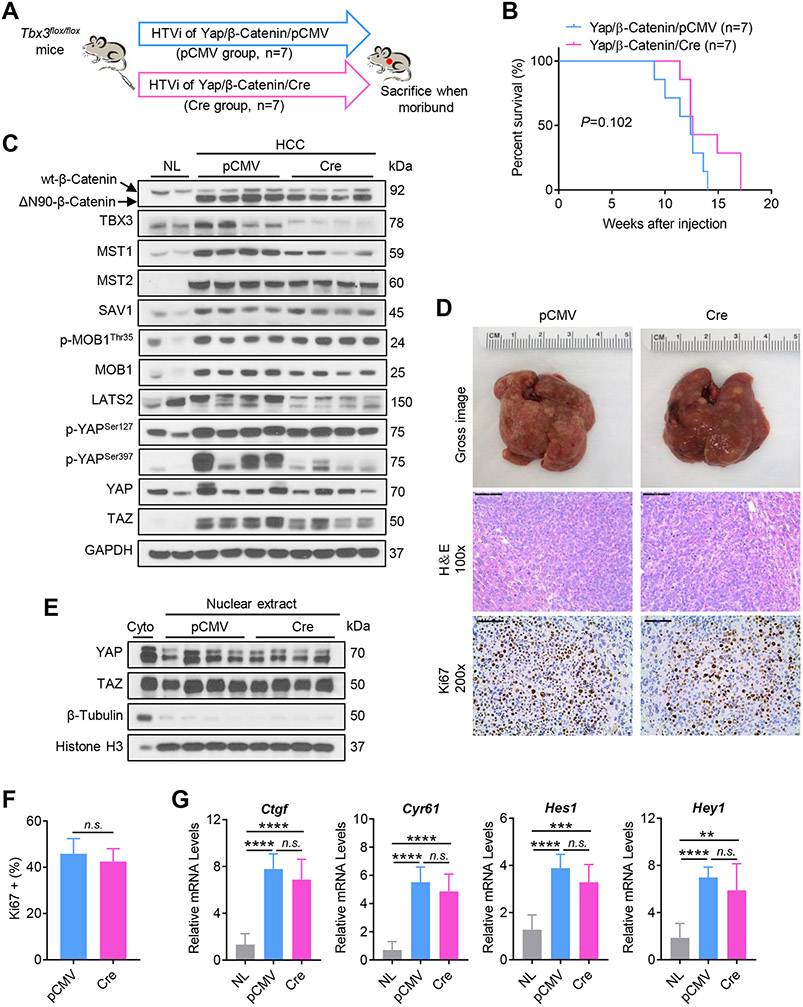
Fig. 4. Ablation of Tbx3 does not accelerate tumor growth in Yap/β-Catenin mice.
(A) Study design. (B) Survival curves of Yap/β-Catenin/pCMV and Yap/β-Catenin/Cre mice. (C) Western blot analysis of lysates from normal livers (NL) and mouse HCCs. GAPDH was used as a loading control. (D) Gross images, H&E staining, and immunohistochemical staining of Ki67 in mouse HCCs. (E) Western blot analysis of nuclear lysates from mouse HCCs. β-Tubulin and Histone H3 were used as loading controls. (F) Quantification of Ki67 positive cells in mouse HCCs. (G) Relative mRNA levels of YAP/TAZ signaling targets (Ctgf, and Cyr61), Notch signaling targets (Hes1, and Hey1) in mouse HCCs. HTVi, hydrodynamic tail vein injection; Scale bar: 200 μm for 100x; 100 μm for 200x. n.s., not significant; **P < 0.01; ***P < 0.001; ****P < 0.0001. (F) Mean ± SD; Unpaired t-test. (G) Mean ± SD; One-way ANOVA test.

