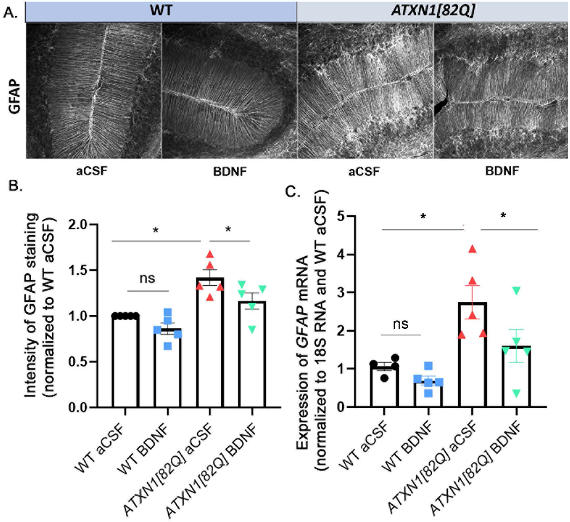
Figure 4. Astrogliosis is reduced with BDNF treatment.

A. Representative images of GFAP staining in the cerebellar molecular and Purkinje cell layers. B. Quantification of GFAP intensity in Bergmann glia. N=5 per group. C. Relative expression of GFAP mRNA in total cerebellar extracts, measured with RTqPCR (normalized to 18S RNA using WT aCSF as reference) N=5 per group. Each data point represent individual mouse, bars show the average with error bars = SEM. * P<0.05, one-way ANOVA followed by Sidak’s post hoc test.
