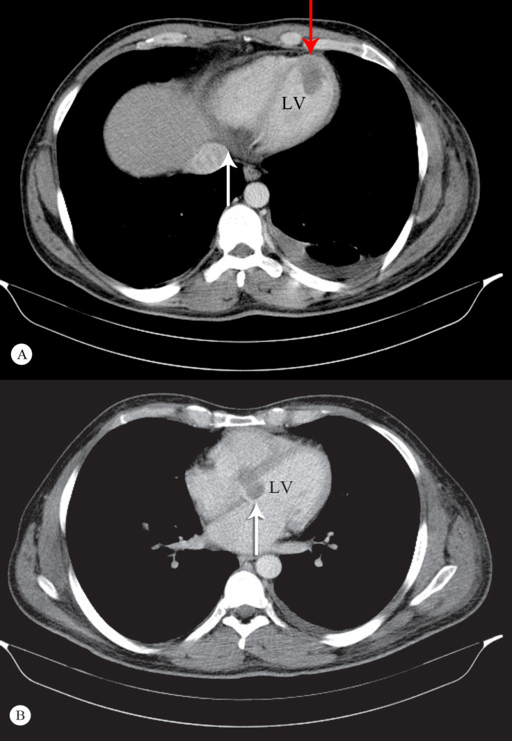
Figure 2.
A) Cross-sectional image of the metastatic involvement of the heart in computed tomography (CT) scanning. Note the infiltration of the basal posterior (and inferior) walls of the left and right ventricular septa (white arrow), as well as the apical protrusion of the tumor (red arrow). B) Cross-sectional CT scan of the heart and the mediastinum at the level of the atria and the main bronchi. Note that the base of the interventricular septum and the mitral annulus is involved (white arrow).

