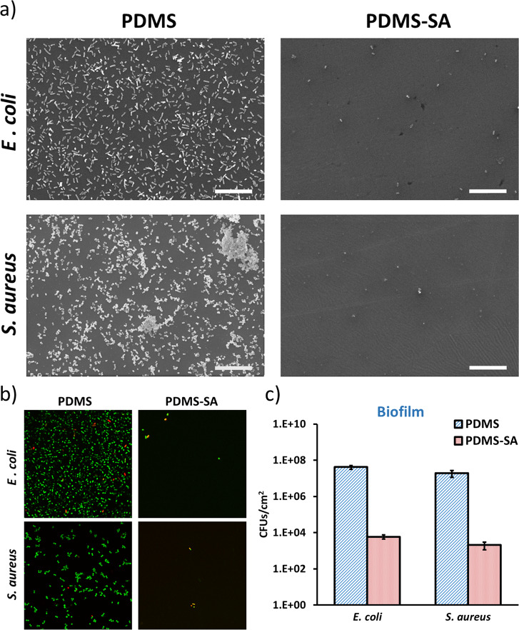
Fig. 8. Antimicrobial activity of PDMS-SA samples against sessile E. coli and S. aureus cells compared to PDMS control samples.
a Representative scanning electron microscopy images of E. coli and S. aureus at the surface of PDMS and PDMS-SA samples after 24 h of incubation (scale bar 20 µm); b Representative confocal fluorescence images of bacteria at the surface of PDMS and PDMS-SA samples after 24 h, as stained by Live/Dead fluorescent labelling, showing live cells in green and dead cells in red/yellow (image size 71 µm × 71 µm); c Inhibition of sessile E. coli and S. aureus cells after 24 h of incubation (inoculum 104 CFU/ml) on PDMS-SA.

