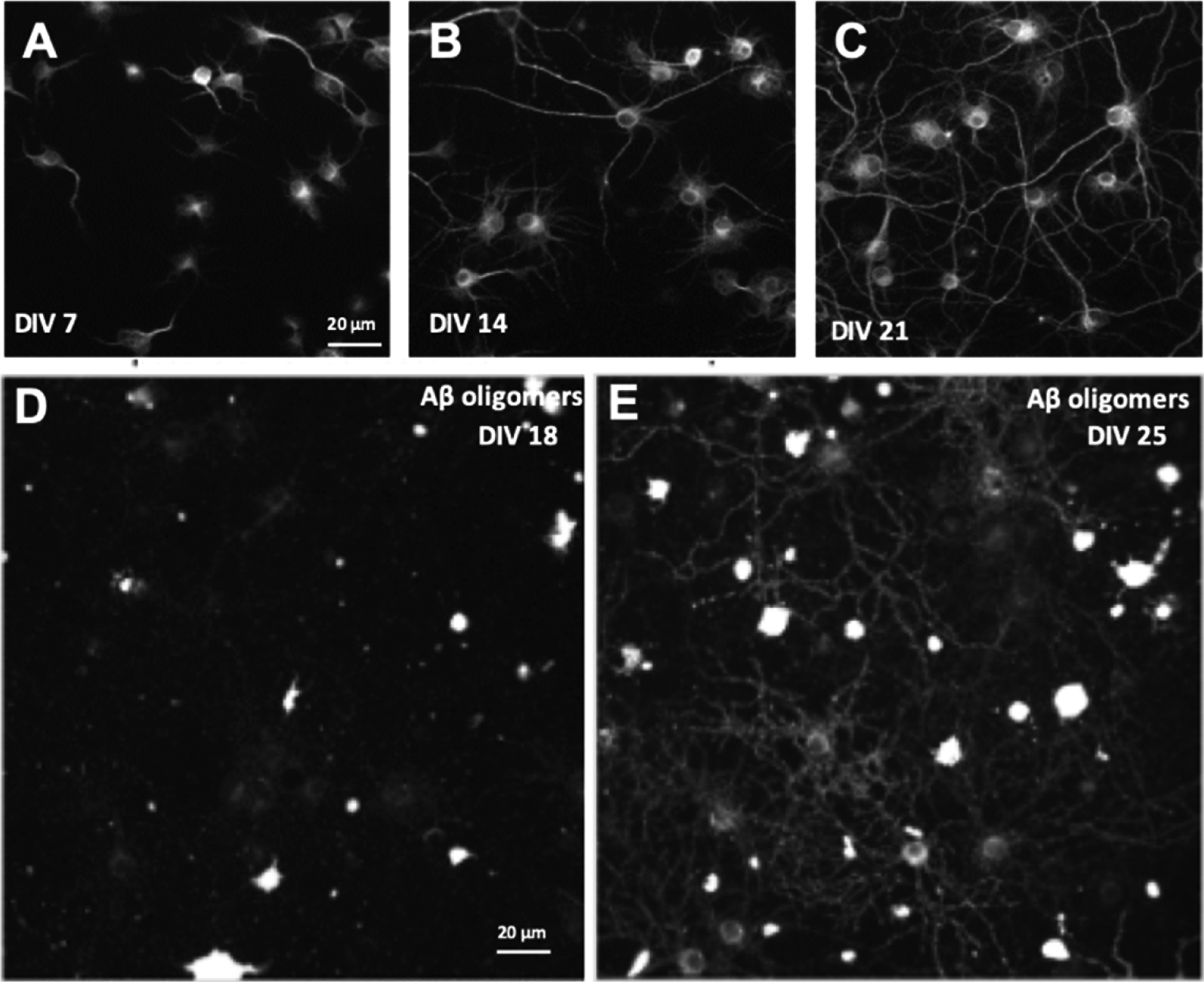
Fig. 2.

Immunofluorescent labeling of primary mixed hippocampal and neocortical neuronal cultures reveals the temporal maturation of neurons and glia in vitro. Cultures that have been maintained for increasing length of time in culture seven days in vitro (DIV7, A), DIV14 (B), and DIV21. (C) cultures were labeled with MAP2 primary antibody and the dendrite segment length quantified via automated image processing. Average dendrite segment length increases over time (DIV7 = 72.7 ± 6.2, DIV14 = 264.2 ± 30, and DIV21 = 529.5 ± 73.8 microns). Cultures were treated with Aβ 1–42 oligomers at DIV18 (D) and DIV25 (E); low binding of oligomers to synaptic receptors was observed at DIV18 but sister cultures treated with oligomers seven days later exhibited increased oligomer binding due to maturation of synaptic oligomer receptor expression (N = 3 independent cell culture preparations). Scale bar (20 μm) for B and C shown in A; scale bar (20 μm) for E shown in D.
