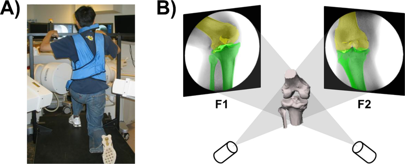
Fig. 1.
(A) The dual fluoroscopic imaging system (DFIS) set up for measurements of knee joint motion during a quasi-static single leg lunge. (B) Illustration of the 3D-2D registration that reproduce the kinematics of the knee in a virtual DFIS by matching the projections of the 3D knee model to the two fluoroscopic images captured along flexion path.

