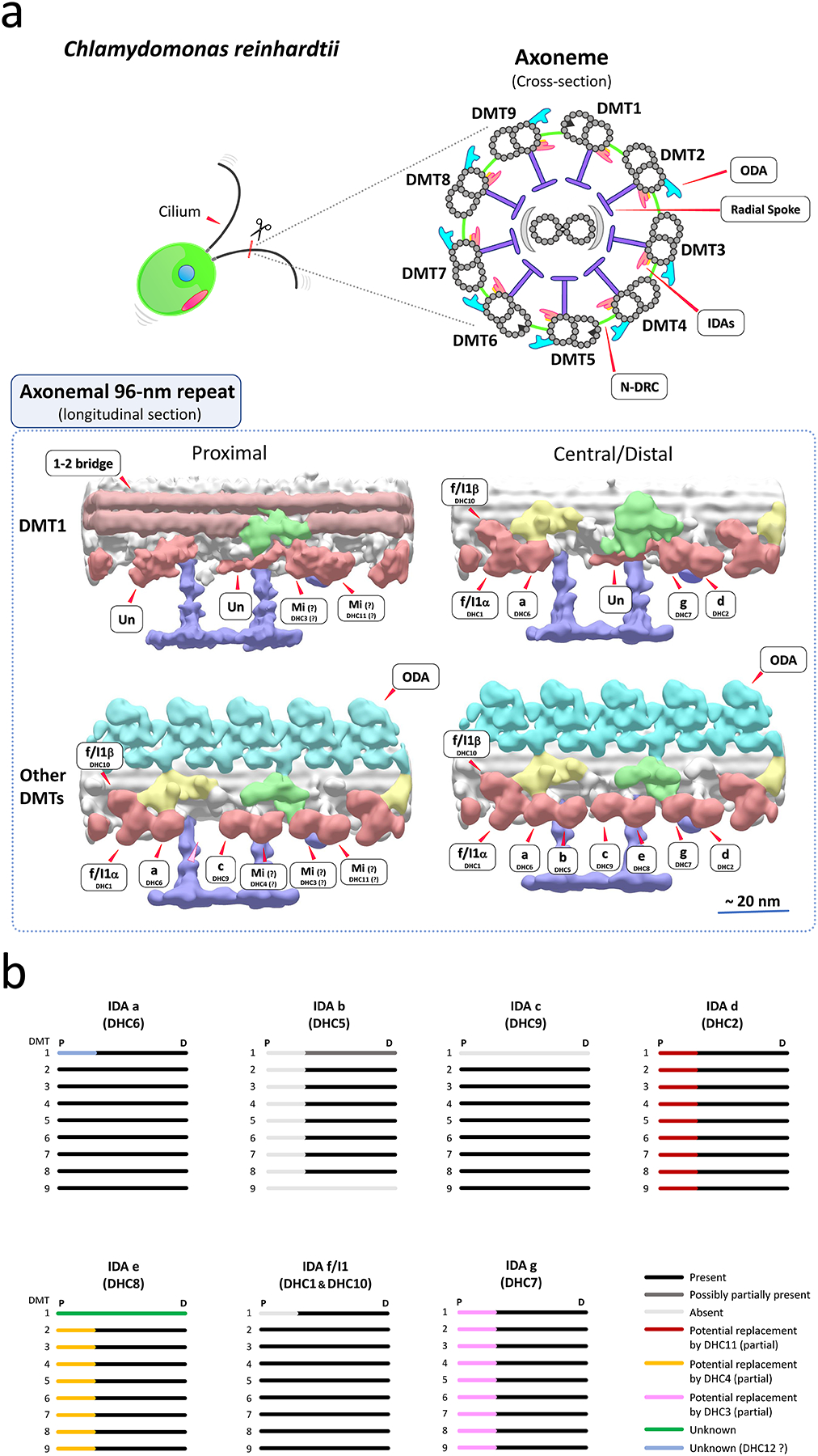
Figure 1. Potential arrangements of IDAs in Chlamydomonas cilia.

(a) (top) Drawings of a Chlamydomonas cell and ciliary axonemal cross-section. (bottom) Hypothesized models of IDA arrangements on surface-rendering images of the 96-nm repeats of Chlamydomonas ciliary doublet microtubules (DMTs). The images are the results of sub-tomogram averages from reconstructed cryo-electron tomograms. Surface-rendering images of the proximal and central/distal regions of DMT1 and other averaged DMTs (DMTs 2–8 or 2–9) are shown. In Chlamydomonas, DMT1 totally lacks ODAs, and also has particularly interesting features including the arrangement of IDAs (Bui et al., 2012; Hoops & Witman, 1983). In the proximal region of ciliary axonemes, the arrangement of IDAs differs from that in the central/distal region, lacking IDA b (DHC5) and possibly minor IDAs replacing some major IDAs (Bui et al., 2012). Approximate locations of ODAs, IDAs, the IC/LC complex of IDA f/I1, N-DRC, radial spokes and the 1–2 bridges are shown in water blue, old-rose red, yellow, green, purple, and light brown, respectively. A light pink arrowhead indicates the missing IDA b location in the proximal portion of the DMT2-9 average. Mi, minor IDA; Un, unknown density likely an IDA species (Bui et al., 2012). The tomograms were reconstructed and published in the previous publication (Bui et al., 2012), and refined for this review. The accession IDs of the density maps (Bui et al., 2012) in EMDataBank (http://www.emdatabank.org/) used to make this figure are as follows: DMT1 proximal region, EMD-2119; DMT1 central region, EMD-2113; DMT2-9 proximal region, EMD-2131; DMT2-8 central/distal region, EMD-2132. (b) A summary of proposed arrangements of IDAs in Chlamydomonas cilia. The figure is adapted/refined from (Bui et al., 2012). Chlamydomonas cilia have heterogeneity in the arrangement of IDAs among DMTs, and this heterogeneity could contribute to generation of proper ciliary waveform. IDA d and e may be partially replaced by minor IDAs DHC11 and DHC4 in the proximal portion of the axonemes (Bui et al., 2012). Also, replacement of IDA g by DHC3 was proposed in (Bustamante-Marin et al., 2020). DMT1 has unique organization of IDAs throughout the axonemes (Bui et al., 2012; Hoops & Witman, 1983). P, Proximal; D, Distal.
