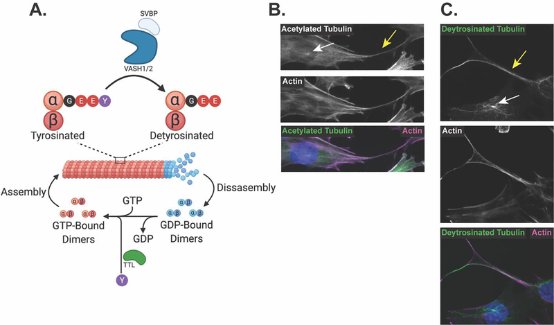
Figure 2: Microtubules are a dynamic structure that become post-translationally modified and are found in osteocytic cell bodies and cell processes.
A. GTP-bound microtubule α/β-tubulin dimers are incorporated into growing filaments. Intrinsic GTP-ase activity of the tubulin dimers cleaves GTP to GDP, promoting disassembly of microtubule filaments, while GTP exchange factors facilitate the transition from GDP- to GTP-bound dimers to promote reassembly of microtubules. VASH1/2 in complex with SVBP removes the terminal tyrosine on α-tubulin in stable microtubules, producing detyrosinated microtubules. This detyrosination can be reversed by TTL, which acts on released α/β-tubulin dimers to ligate the terminal tyrosine onto the α-tubulin tail. B. Immunofluorescent staining of acetylated tubulin and actin microfilaments shows their localization to both the cell body, primary cilia (white arrow) and the dendrite-like osteocyte cell processes (yellow arrow) of Ocy454 cells. C. Immunofluorescent staining of detyrosinated tubulin and actin microfilaments shows their localization to the cell body, primary cilia (white arrow) and the dendrite-like osteocyte cell processes (yellow arrow) of Ocy454 cells.

