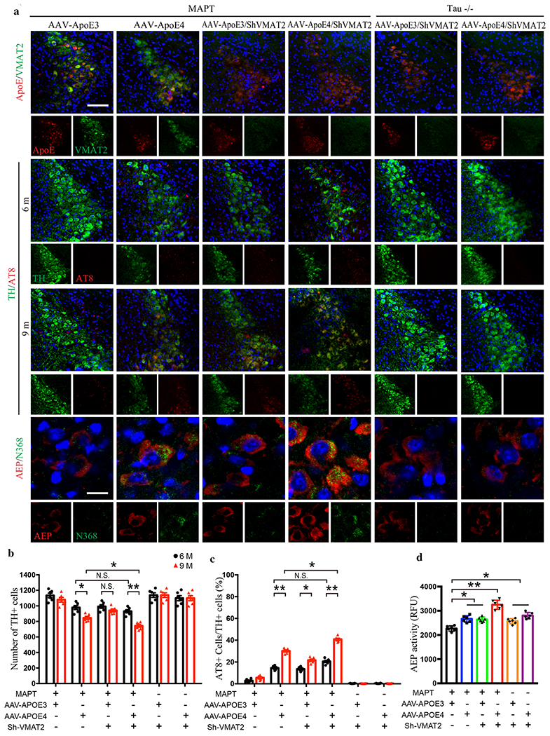
Fig. 6. Deletion of VMAT2 aggravates ApoE4-triggered AD pathology and LC degeneration.

AAV-ApoE3/ApoE4 and Lenti-Sh-VMAT2 were injected into the LC of MAPT or Tau −/− mice, and then mice were assessed for Tau pathology and neuronal loss 3 months and 6 months later (6 and 9-month-old group, respectively), a. The upper panels are the representative images of ApoE and VMAT2 immunofluorescence co-staining verifying viral infection of AAV-ApoE3/ApoE4 and Lenti-Sh-VMAT2. Representative images of immunofluorescence staining of TH (green), AT8 (red), and DAPI (blue) in 2nd panel (6 month) and 3rd panel (9 month). The last panel’s representative images are AEP (red) and Tau N368 (green) immunofluorescence co-staining to show AEP activation and Tau cleavage in the LC. Scale bar is 100 μm. b. Stereological cell counting of TH+ cells in the LC region showed neuronal loss mediated by ApoE4 overexpression and VMAT2 deficiency, c. Quantification of AT8+ cells in TH+ cells in the LC to show Tau phosphorylation, d. AEP enzymatic assay in the LC for AAV-ApoE3/ApoE4 and Lenti-Sh-VMAT2-injected MAPT or Tau −/− mice. All data in b-d were analyzed using two-way ANOVA and shown as mean ± SEM. N=6 per group. * <0.05, ** <0.01.
