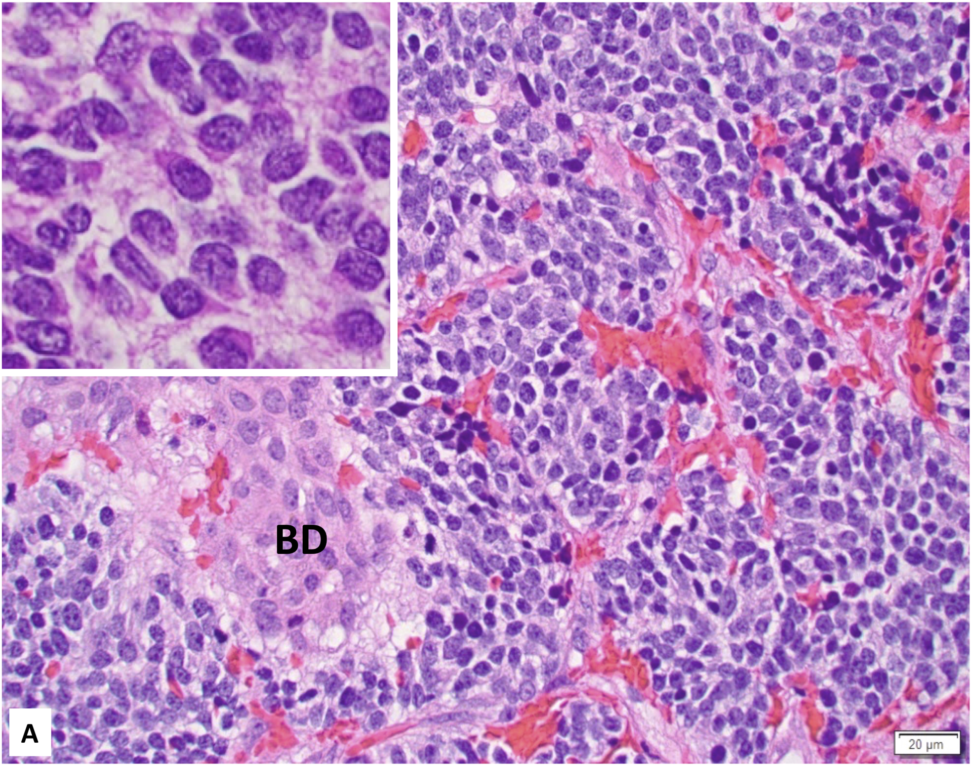
Figure 1:

A. Favorable Histology Stage 4S Neuroblastoma (Poorly differentiated subtype with a low MKI) involving the liver (Inset: Higher magnification demonstrating “Salt-and-Pepper” Nuclei). BD: bile duct; B. Unfavorable Histology Stage 4S Neuroblastoma (Poorly differentiated subtype with a high MKI) involving the liver (Inset: Higher magnification demonstrating prominent nucleolar formation - nucleolar hypertrophy).
