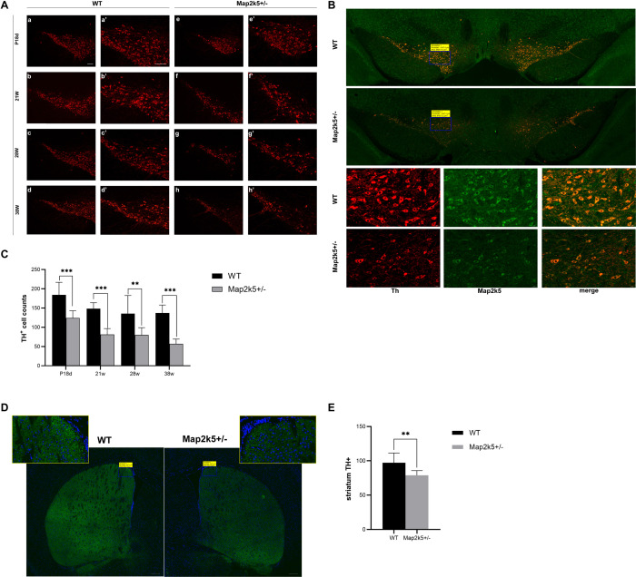
FIGURE 4.
(A) Coronal sections of substantia nigra pars compacta (SNc) were prepared from P18d, 21, 28, and 38 weeks and immunostained with an anti-TH antibody. Map2k5+/– mutant mice had decreased TH+ cell number in SNc at P18d, 21, 28, and 32 weeks. (a–d) Immunostained TH+ cells in SNc of Map2k5+/+ WT mice (100x). (e–h) TH+ cells in SNc of Map2k5+/– mutant mice (100×). (a’–h’) High-magnification images (200 ×) of the corresponding section in (a–h). Scale bar, 100 μm. (B) TH+ and Map2k5+-stained cells, and merged pictures of Map2k5+/– mutant and WT mice in SNc of at 38 weeks. Low-magnification images (50×) and high-magnification images (400×). 200 μm (50×), 20 μm (400×). (C) TH+ cell counts in SNc were reduced in female Map2k5+/– mutant mice compared with WT mice at P18d, 21, 28, and 38 weeks (P18d < 0.001, P21w < 0.001, P28w = 0.005, P38w < 0.001). (D) Coronal sections of striatum immunostained with anti-TH antibody. We found decrease immunofluorescence in Map2k5+/– mutant mice compared with WT mice. Low-magnification images (50 ×) and high-magnification images (630×). Scale bar, 200 μm (50×), 20 μm (630×). (E) Mean immunofluorescence intensity of each slice was analyzed. There was decreased TH density in Map2k5+/– mutant mice (P = 0.006). Results are plotted as mean with SEM. Bars represent means with SEM. **P < 0.01, ***P < 0.001.

