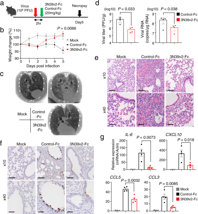
Fig. 5. Therapeutic efficiency of 3N39v2-Fc in a COVID-19 Hamster Model.
a Schematic overview of the animal experiment. b Percent body weight change was calculated from day 0 for all hamsters. c Axial CT images of the thorax 5 days after infection. d Quantification of plaque-forming units (PFU) from lung homogenates and genomic SARS-CoV-2 RNA as copies per μg of cellular transcripts. e H&E staining and f SARS-CoV-2 antigen staining of hamster lung lobes. Scale bars, 100 μm (upper panel) and 40 μm (lower panel). g mRNA expression of inflammatory or chemotactic cytokines in hamster lung lobes. b, d, g Data are mean ± SEM of n = 6 for mock group, 4 for each treated group. P-values by two-sided unpaired t-test.

