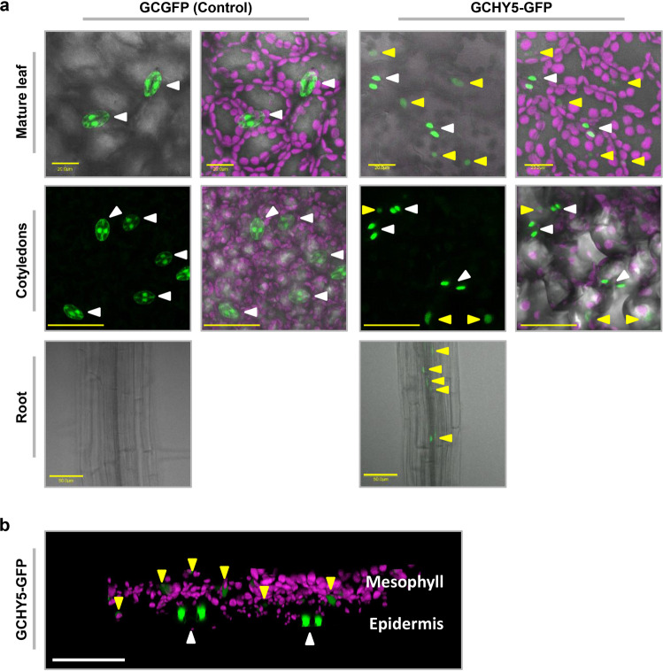
Fig. 7. HY5 produced within guard cells is translocated to mesophyll and root cells.
a Distribution of GCGFP (control) or GCHY5-GFP in mature leaves, cotyledons and roots. All panels are merged images of white-light, chlorophyll-autofluorescence (stained magenta), and GFP-fluorescence (stained green). Scale bars (yellow) are 20 µm for mature leaves and 50 µm for cotyledons and roots. b 3D simulation providing side views of GCHY5-GFP cotyledons, composed of epidermis and mesophyll cell layers. Image is a merge of chlorophyll-autofluorescence (stained magenta), and GFP-fluorescence (stained green). Bar = 50 µm. a, b White arrows indicate the location of GFP in guard cells and yellow arrows indicate the location of GFP in mesophyll cells of mature leaves and the cotyledons, and within the phloem of the roots.

