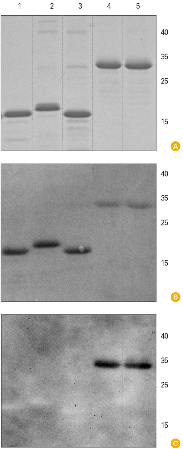
Fig. 2. Characterization of rotavirus A (RVA) recombinant antigens. Lanes: VP8*-[P8] (1), VP8*-[P6] (2), VP8*-[P4] (3), ep-875 (4), VP5* (5). (A) Electrophoresis analysis, 8%–20% SDS-PAGE (sodium dodecyl sulphate–polyacrylamide gel electrophoresis), staining with Coomassie G-250. (B) Western blot membrane stained by Ponceau S. (C) Western blot analysis with commercial polyclonal antisera to 5 RVA strains: Wa-G1P[8], Ds-1 G2P[4], Ito-G3P[8], HOCHI-G4P[8], 69M-G8P[10] (Cat# MBS316568; MyBioSource Inc., San Diego, CA, USA) as primary antibodies and secondary anti-species antibodies conjugated with horseradish peroxidase. Positions of molecular weights (kDa) markers are indicated in the right.