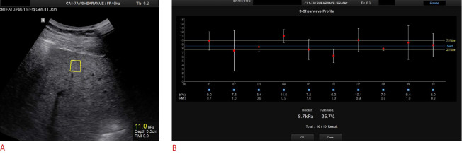
Fig. 2. Point shear-wave elastography in a 44-year-old man with significant fibrosis.
A. Intercostal ultrasound image in the supine position shows a region of interest located under the liver capsule at a depth of 3.5 cm. B. Ten valid measurements are reported with a final median liver stiffness value of 8.7 kPa indicated in the lowest part of the image. A good interquartile range is also shown.

