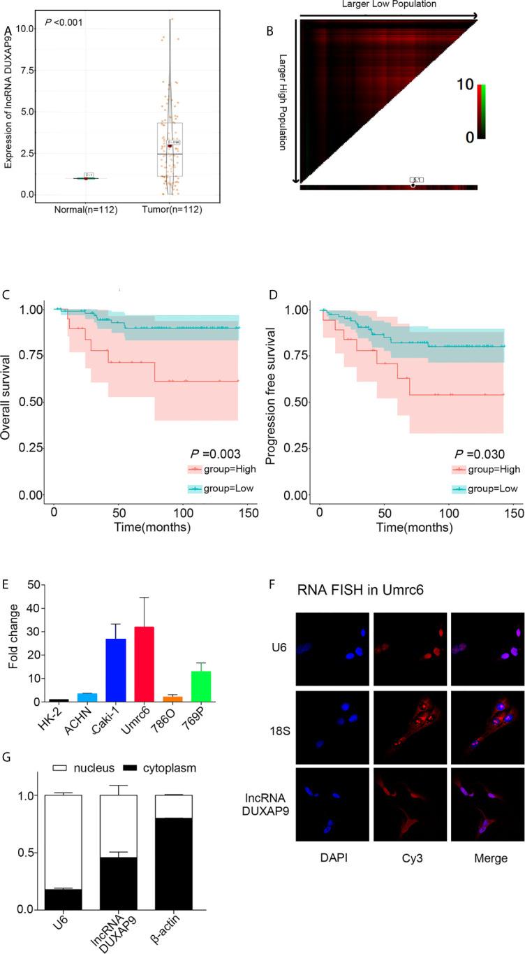
Figure 1.

DUXAP9 was upregulated in renal cancer and predicted poor prognosis in localized ccRCC patients. (A) Relative DUXAP9 expression levels in 112 pairs of localized ccRCC and matched adjacent non-tumor tissues from the SYSUCC Biobank, as detected by qRT-PCR. (B) Optimal cutoff to divide ccRCC patients into high and low DUXAP9 expression groups, as determined by X-tile bioinformatics software based on the integral optic density. (C, D) Kaplan–Meier curves showing that high DUXAP9 expression (n=19) in localized ccRCC tissues was significantly associated with poor OS and PFS rates, compared with low expression (n=93). (E) Relative DUXAP9 expression levels in renal cancer cell lines, as detected by qRT-PCR. (F) FISH analysis of the subcellular distribution of DUXAP9 in Umrc6. (G) Subcellular fractionation and qRT-PCR analyses of DUXAP9 expression in the nucleus and cytoplasm. Data represent mean ± SD from three independent experiments. ccRCC, clear cell renal cell carcinoma; qRT-PCR, quantitative real-time PCR; OS, overall survival; PFS, progression-free survival; FISH, fluorescence in situ hybridization.
