FIGURE 4.

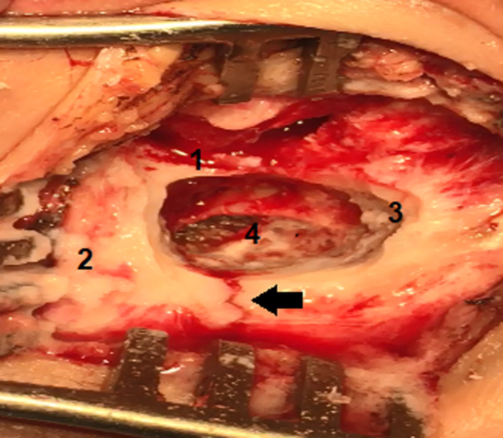
Transoperative View of the right side: 1‐Posterior bony of external ear canal. 2‐mastoid tip. 3‐Tegmen tympani. 4‐Mastoid cavity after drilling had been done by a surgeon. Black Arrow‐Fracture line

Transoperative View of the right side: 1‐Posterior bony of external ear canal. 2‐mastoid tip. 3‐Tegmen tympani. 4‐Mastoid cavity after drilling had been done by a surgeon. Black Arrow‐Fracture line