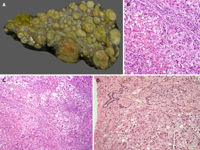
Figure 1.
Metabolic liver disease. A: Explant liver specimen of a case of tyrosinemia with nodules of varying sizes; B: Tyrosinemia liver with steatosis [hematoxylin and eosin (HE staining)]; C: Tyrosinemia liver with cirrhosis, hepatocellular ballooning, and fatty change (HE staining); D: Galactosemia with bridging fibrosis, bile ductular reaction, fatty change and rosetting (HE staining).

