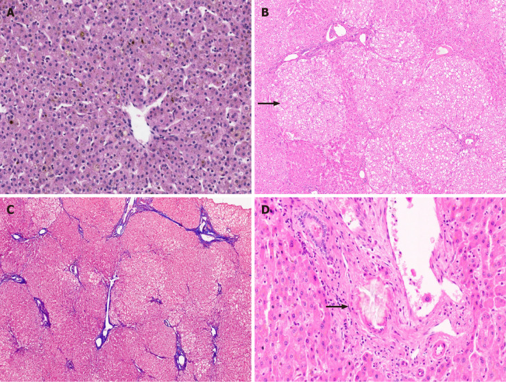
Figure 5.
Metabolic liver disease. A: Canalicular bilirubinostasis in case with urea cycle defect [hematoxylin and eosin (HE staining)]; B: Explanted liver in Arginase-1 (ARG-1) deficiency displaying nodules of pale enlarged hepatocytes (arrow, HE staining, B); C: Focal bridging fibrosis in a case of ARG-1 deficiency [Masson trichrome staining]; D: Portal vessel with oxalate crystals in primary hyperoxaluria type 1 (arrow, HE staining).

