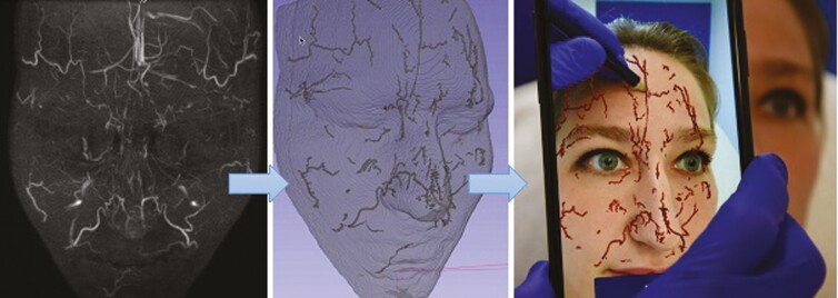
Figure 7.
Magnetic resonance angiography findings and augmented reality (AR) visualization in a 26-year-old female. (A) Infrared thermally enhanced MRI without contrast. Frontal view of a maximum intensity projection (MIP) from the 3-dimensional time-of-flight multiple overlapping thin slab acquisition (3D-TOF MOTSA). (B) Segmentation of subcutaneous arteries. 3D image after isolation of the arteries and conversion to a 3D volume. (C) Visualization of subcutaneous arteries through AR and projection on the face of the patient with a smartphone camera.

