
Figure 3. Functional upregulation of delta opioid receptors (DORs) in the spinal cord of rats with neuropathic pain.
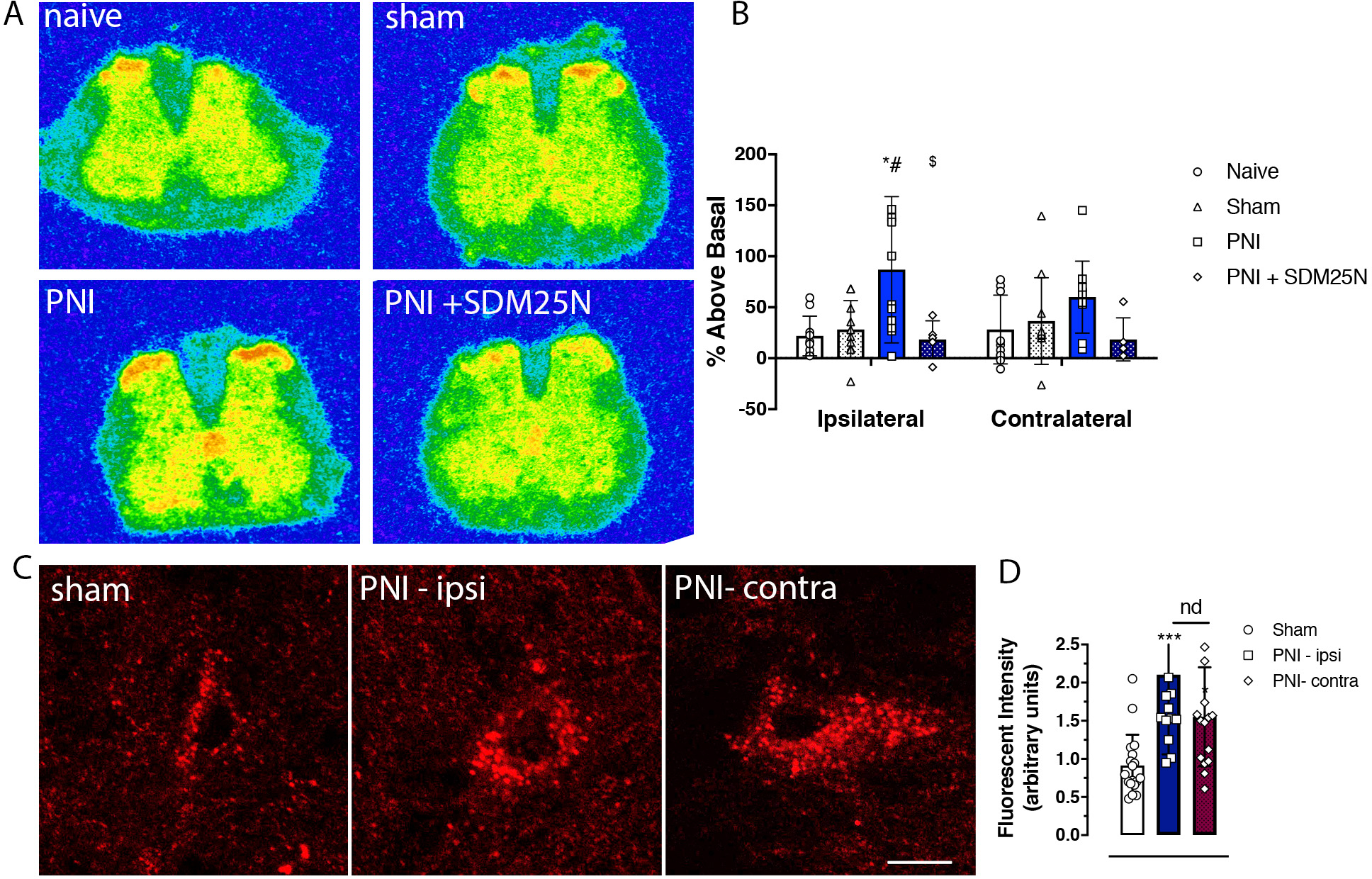
A. Representative autoradiograms of DOR agonist-induced [35S]GTPγs binding in transverse lumbar spinal cord sections from pain naïve, sham, and peripheral nerve injury (PNI) rats. B. Increases in DOR agonist deltorphin (DLT; 1 uM)-stimulated [35S]GTPγs binding are evident in the superficial dorsal spinal cord of PNI mice compared to both pain-naïve and sham controls. This increase in agonist-induced GTP binding was prevented by co-incubation of the DOR antagonist SDM25N (1 uM). C. High magnification confocal microscopic images of fluorescent-deltorphin (Fluo-DLT)-labeled neurons in the rat dorsal spinal cord 20 min after intrathecal administration of the fluorescent agonist (0.8 nmol). Images are presented in glow scale, where white represents the highest fluorescence intensity and red represents the lowest (black indicates the absence of fluorescent signal). Florescent puncta are evident throughout the perikaryal cytoplasm, where greater fluorescence is evident in spinal cord sections of PNI (14 days post-surgery) compared to sham control rats. Scale bar is 10 μm. D. Densitometric quantification of fluorescent labeling showing a significant bilateral increase of Fluo-DLT internalization in PNI compared to sham surgery control rats; there was no difference between the ipsilateral and contralateral sides of the dorsal spinal cord of PNI rats.