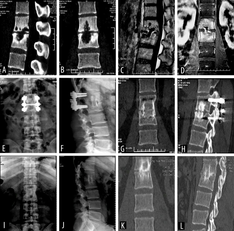
Figure 3.
A 30-year-old male patient diagnosed with L1–2 vertebral tuberculosis was treated with posterior affected-vertebrae fixation, anterior subdiaphragmatic extraperitoneal approach for thorough lesion removal, and autologous iliac bone graft fusion. (A, B) Preoperative computed tomography (CT) showed obvious bone destruction. (C, D) Enhanced magnetic resonance imaging before surgery showed vertebral signal changes, vertebral bone destruction, and paravertebral abscess. (E–H) X-ray and CT at 6 months after surgery showed pedicle screw fixation and good bone graft fusion. (I–L) X-ray and CT 5 years after surgery showed that the pedicle screw had been completely removed, the lesion had completely cured, and the bone graft had completely fused.

