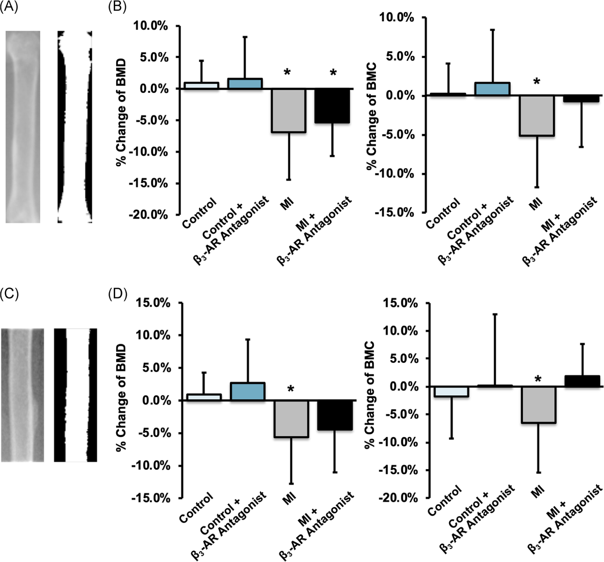
FIGURE 5.

(A,C) X-ray and thresholded images of the whole femur (A) and the femoral diaphysis (C). (B,D) The average change of femoral BMD (left) and BMC (right) from baseline to 9 days post-MI for the whole femur and femoral diaphysis. Both BMD and BMC decreased significantly in MI mice, and this decrease was greater for untreated mice than for β3-AR antagonist treated mice. * denotes p ≤ .05 between baseline and 9 days post-MI. β3-AR, beta 3-adrenergic receptor; BMC, bone mineral content; BMD, bone mineral density; MI, myocardial infarction
