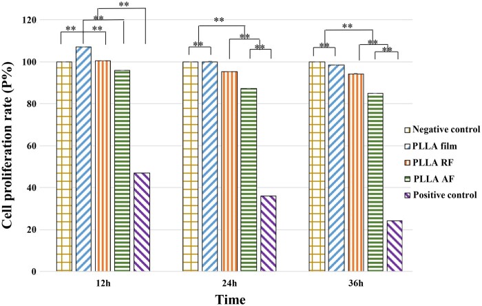
Figure 2.
The proliferation rate of the PC12 cells on the three PLLA substrates after 12, 24 and 36 h. The negative control cells were cultured in the differentiation medium alone; the positive control cells were cultured in the medium plus 0.7% acrylamide solution without any biomaterial. All the data were presented as mean ± standard deviation. **P < 0.01.

