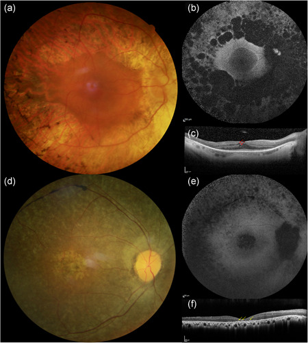
Figure 3.

Clinical phenotype of a CNGB1‐related retinitis pigmentosa patient (F3791‐CIC06919). Fundus photographs of the right (a) and left (d) eyes show a waxy optic disc, narrow vessels and peripheral bone spicules. On short‐wavelength fundus autofluorescence (b, e), the central area of preserved tissue is surrounded by a ring of increased autofluorescence that demarks the limits of the peripheral atrophy. On optical coherence tomography (c, e) all retinal layers look centrally well preserved, while peripherally a thinning of the outer layers is evident
