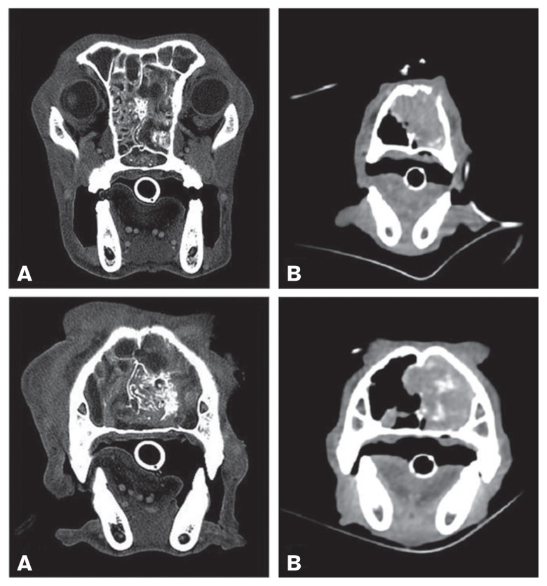
Figure 3.
Transverse CT images (A — Case 1, B — Case 15) of the bone and soft tissue window. In all images, a mass involving the nasal cavity (A and B left) is clearly visible, characterized by patchy contrast enhancement (B) and destruction of turbinates and parts of the bony nasal septum (A and B). Partial lysis of the nasal bone is also present (B). The mass extends into the choanae and the rostral aspect of the nasopharynx (A).

