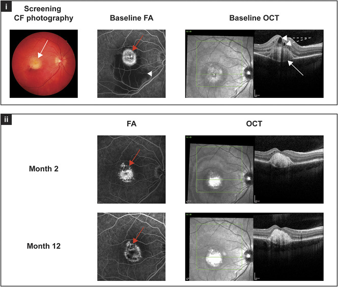
Fig. 1.
Image studies from “Patient A” at screening/baseline, and Months 2 and 12 (Table 1): (i) at baseline, the typical yellowish, egg-yolk–like, round and isolated lesion in the center of the macula is visible in the color fundus photograph (white arrow). The FA at baseline shows an active CNV with leakage (red arrow) as well as subretinal hemorrhage in the inferior part of the macula (white arrowhead). Optical coherence tomography scans at baseline reveal intraretinal and subretinal fluid (white arrows with broken line) as well as subretinal vitelliform material (white arrow). (ii) Fluorescein angiography images at Months 2 and 12 show a quiescent lesion with only staining (red arrow), and the subretinal hemorrhage has resolved. Optical coherence tomography scans at Month 2 show no subretinal fluid, the intraretinal fluid is improved, the foveal pit is visible, and subretinal hyperreflective material is present. At Month 12, the foveal structure is unchanged, there are some intraretinal cystoid changes, but no signs of CNV activity are visible on OCT scans.

