
FIGURE 1.
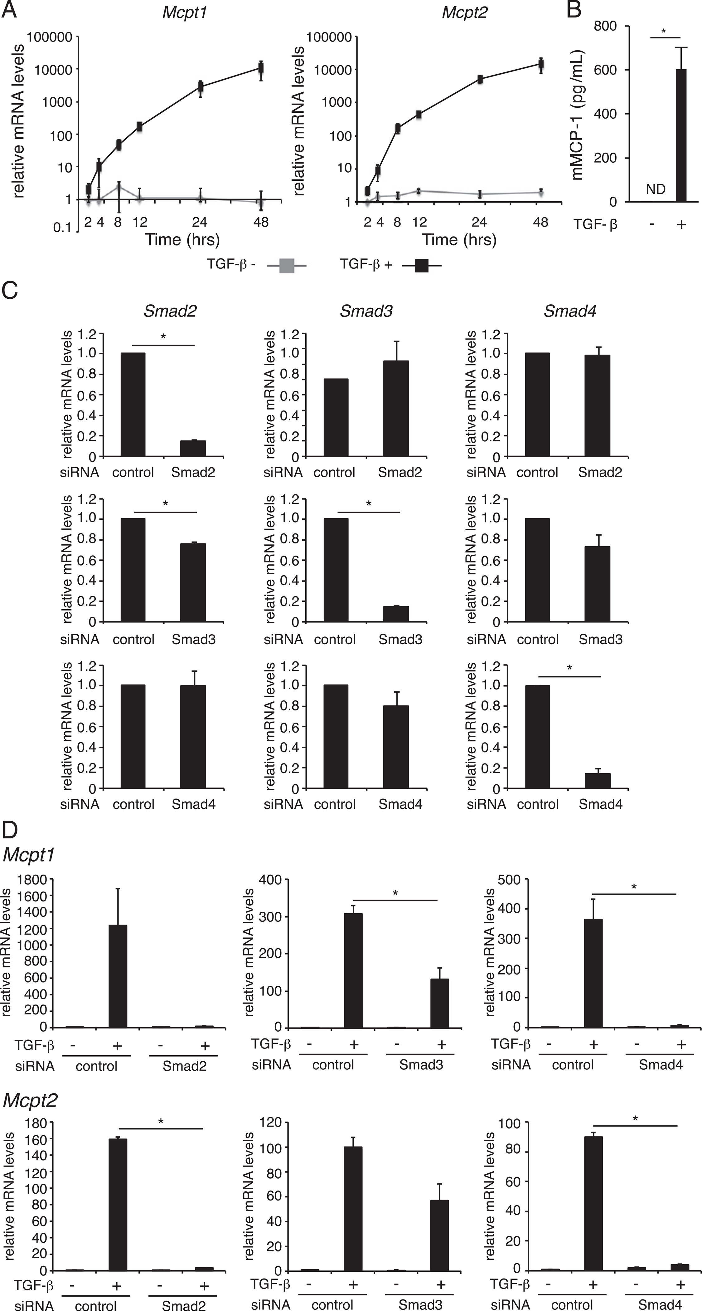
Effect of TGF-β signaling on the expression of Mcpt1 and Mcpt2 in BMMCs. (A) BMMCs were treated with 1 ng/ml TGF-β for the indicated times. The mRNA expression levels of Mcpt1 and Mcpt2 were assessed by quantitative RT-PCR. (B) mMCP-1 protein concentrations in culture media of BMMCs. BMMCs were cultured in the presence of 1 ng/ml TGF-β for 72 h. The culture media of BMMCs (1.0 × 10⁶ cells/500 μl), which were stimulated with 1 m-M A23187 for 1 h, were harvested to determine mMCP-1 protein concentrations by an ELISA. (C) The mRNA levels of Smad2, Smad3, and Smad4 in siRNA-transfected BMMCs at 48 h after siRNA transfection. (D) The mRNA levels of Mcpt1 and Mcpt2 in siRNA-transfected BMMCs at 8 h after addition of TGF-β. The expression of each mRNA was normalized to that of GAPDH mRNA by calculation of the cycle threshold values. The data are presented as the mean ± SD of three independent experiments performed with triplicate samples (A, C, and D). A typical result of two independent experiments performed with triplicate samples is shown (B). *p < 0.05.