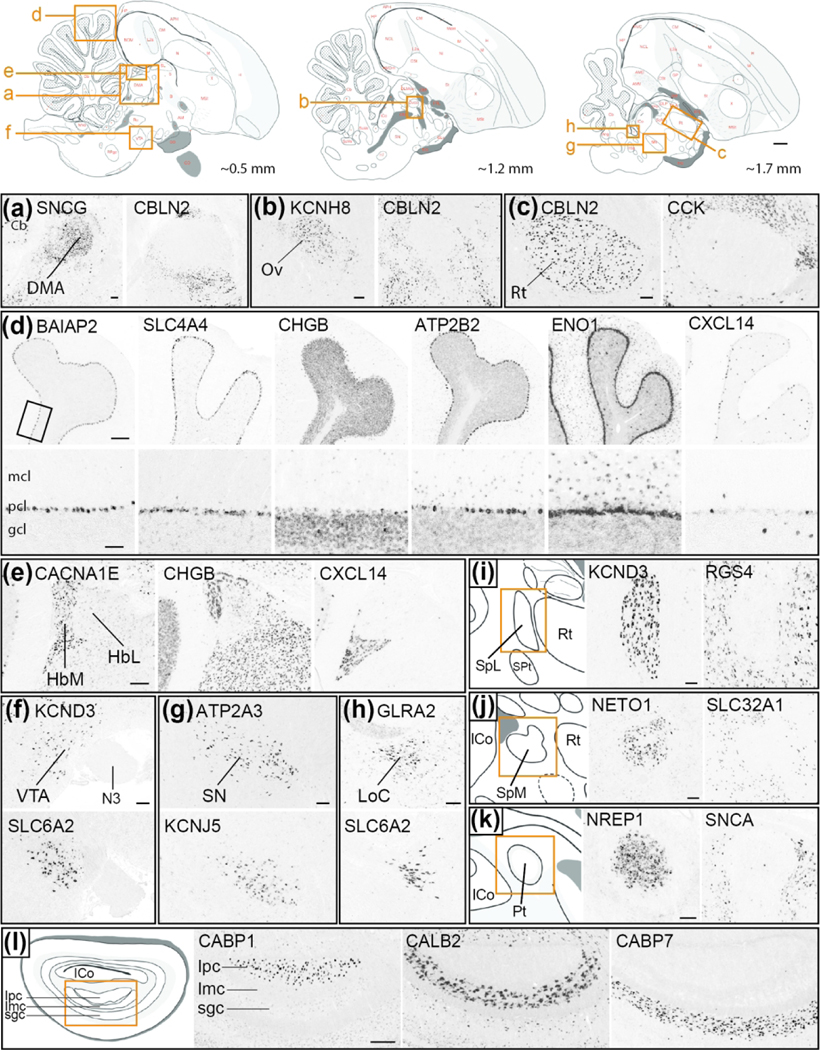
Figure 15. Comparative Neuroanatomy genes: diencephalon and brainstem (Portal 4).
Top row: Schematic drawings of parasagittal sections of the adult male zebra finch brain depict major brain subdivisions and specific nuclei shown in this figure with the exception of the brain areas shown in panels I-L. Red letters and dots indicate structure annotations as they appear in ZEBrA, vermillion rectangles indicate the position of the images shown in other panels, and numbers at bottom right of each diagram provide position (in mm) relative to the midline. Other panels: Examples of in situ hybridization images of genes that show differential expression in various parts of the diencephalon and brainstem, taken from the Bird Brain Markers (sub-portal 4A). (a-c) Positive/negative markers of thalamic nuclei DMA (SNCG/CBLN2), Ov (KCNH8/CBLN2) and Rt (CBLN2/CCK). (d) Markers of cerebellar cortical layers show distinctly differential expression in pcl (BAIAP2, SLC4A4), gcl (CHGB), pcl/mcl (ATP2B2, ENO1), or in sparse cells in gcl (CXCL14, among other patterns; the bottom panels show high magnification views of interface zone across layers, taken from region depicted by rectangle in top left panel. (e) Markers that delineate the habenula and/or differentiate its medial and lateral subdivisions. (f-h) Markers of VTA (KCND3, SLC6A2), SN (ATP2A3, KCNJ5) and LoC (GLRA2, SLC6A2) in brainstem tegmentum. (i-k) Positive/negative markers of pre-tectal nuclei SpL (KCND1/RGS4), SpM (NETO1/SLC32A1) and Pt (NREP1/SNCA). (l) Markers of ipc (CABP1), imc (CALB2) and sgc (CABP7) in the optic tectum. Abbreviations: Cb, cerebellum; DMA, dorsomedial nucleus of the anterior thalamus; gcl, granule cell layer of Cb; HbL, lateral habenular nucleus; HbM, medial habenular nucleus; Imc, magnocellular part of the isthmic nucleus; Ipc, parvocellular part of the isthmic nucleus; LoC, locus coeruleus; mcl, molecular layer of Cb; N3, root of third cranial nerve; Ov, nucleus ovoidalis; pcl, Purkinje cell layer of Cb; Rt, nucleus rotundus; SN, substantia nigra; sgc, stratum griseum centrale of the optic tectum; SpL, nucleus spiriformis lateralis; SpM, nucleus spiriformis medialis; VTA, ventral tegmental area. For other abbreviations in top row diagrams, please consult the histological atlas in ZEBrA. Scale bars: top row diagrams = 1 mm; (a-c) = 200 μm; (d): low-resolution panels = 200 μm, high-resolution panels = 100 μm; (e-k) = 200 μm; (l) = 400 μm.

