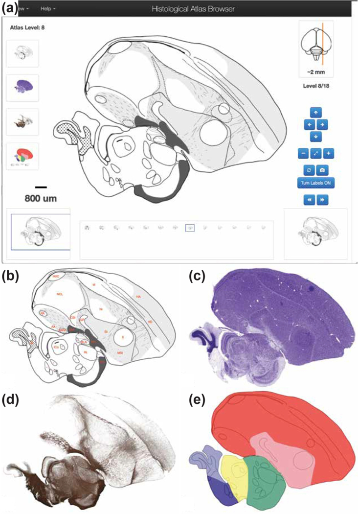
Figure 6. The Histological Atlas Browser.
(a) View of the Browser depicting an example of a drawing from one selected level of the Histological Atlas. The full set of parasagittal levels currently available is shown as thumbnails at the bottom of the browser. The thumbnail icons on the left allow users to toggle across each of the four images types. The position of the section being viewed relative to the midline is illustrated by the orange line on the dorsal-view schematic drawing of the brain, in the top right corner of the bowser. The distance (in mm) of the current section from the midline, as well as the section level in the series, are indicated beneath that schematic. The navigation buttons on the lower right provide options for panning (arrows) or zooming (+ and −) an image, resetting the image zoom level (diagonally opposing arrows), toggling across the four image types for a selected level (circular arrows), requesting an image from ZEBrA (camera symbol), turning anatomical labels ON/OFF, and moving to the next or previous image in the series (>> or <<). The Pan function can also be applied by clicking and dragging the main image in any desired direction, and all other navigation functions can be activated by the appropriate keyboard stroke, as described under the “Navigation Help” tab in the upper-left corner of the browser. The size and position of the field of view shown in the browser is indicated by the blue rectangle over the icon on the bottom left, and the image scale is indicated above that icon. (b) View of the same example drawing as in (a), with Turn Labels button activated (ON position). (c-e) Views of the Nissl- and myelin-stained sections, and of the colorized drawing depicting the: pallium (red), sub-pallium (pink), diencephalon (green), midbrain (yellow), pons (dark blue) and cerebellum (light blue).

