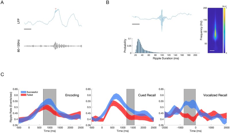
Figure 3.
Hippocampal ripples during encoding and recall predict successful associative memory. Ripple events were detected using a bipolar montage from the electrodes located in or closest to the hippocampus, using a previously published method (80–120 Hz, 20–200 ms duration).10 To reduce the detections of pathological high frequency oscillations (HFOs), detections were restricted to regions outside of SOZ, and with trials which did not contain an IED. (A) Sample of a raw EEG tracing (blue) with detected ripple event (red arrow), with bandpass filtered (80–120 Hz) tracing (black). Scale bar = 125 ms (B) Characteristics of all detected hippocampal ripples. An average of 1955 ripple events were detected per patient across all conditions. Top: Grand averaged ripple response (left) and spectrogram (right, 10–200 Hz) demonstrates a peak frequency between 80 Hz and 100 Hz. Scale bars = 125 ms. Bottom: Histogram showing detected ripple duration, which follows skewed log distribution. Mean ripple duration 39.8 ms, SD 18.3 ms. (C) Average ripple rate between Successful and Failed Associative Memory Trials (mean ± standard error of the mean). Top: Successful encoding is characterized by a higher hippocampal ripple rate (blue) compared to failed encoding (red) between 750 ms and1375 ms after stimulus presentation (grey box, n = 14, P < 0.05, cluster-corrected). Middle: Successful cued recall is characterized by a higher ripple rate compared to failed cued recall between +1250 ms and +1625 ms after stimulus presentation (grey box, n = 14, P < 0.05, cluster-corrected). Bottom: Successful cued recall is characterized by a higher ripple rate from −750 ms to 0 ms aligned voice response (grey box, n = 9, P < 0.05, cluster-corrected).

