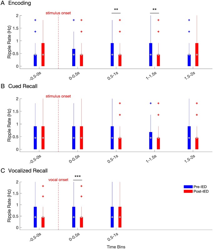
Figure 5.
Hippocampal ripple rate in the pre and post-IED window during encoding and recall. Ripple rates across all hippocampal electrodes in 500 ms pre- (blue) versus 500 ms post-IED (red), binned by time of detected IEDs (500-ms bin windows) for IEDs detected during (A) encoding, (B) cued recall, and (C) voice-aligned recall. Box and whisker plots represent the median (circles) and IQR (bars), along with extreme values (whiskers and outliers). A reduction in ripple rate after an IED event was found during the 0.5–1 s (Z = 2.8838, nIEDs = 209, P = 0.004) and 1–1.5 s (Z = 3.0873, nIEDs=266, P = 0.002) windows during encoding. In addition, ripple rates were significantly reduced in the time window during (0–0.5 s) vocalization during cued recall (Z = 3.9, nIEDs = 177, P < 0.001).

