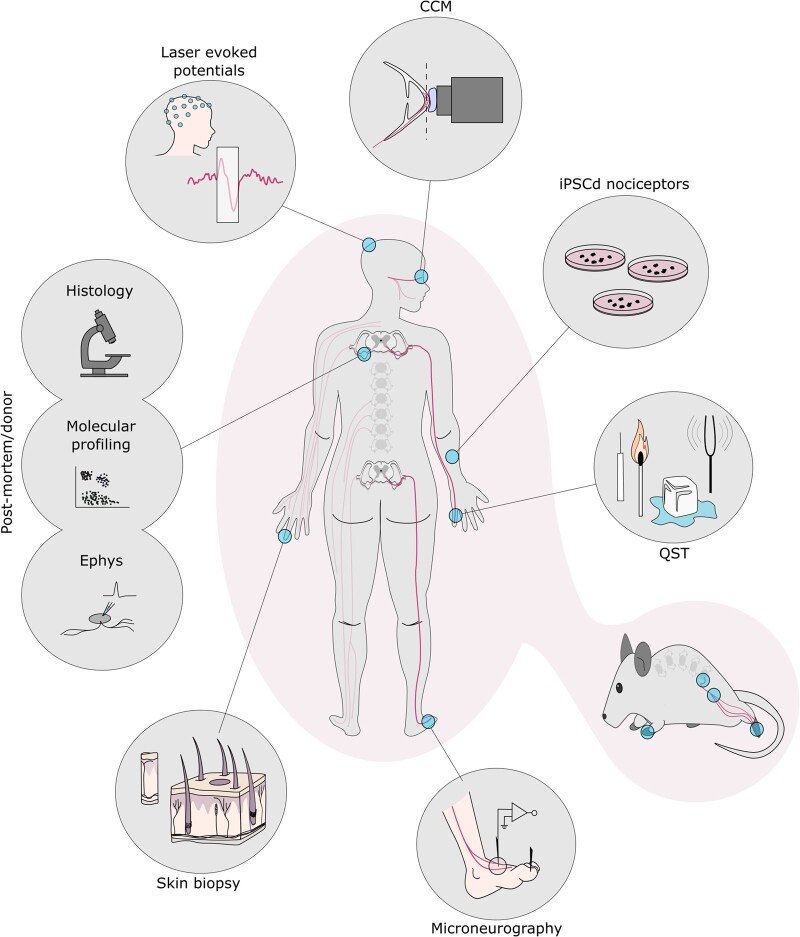
Figure 1.
Highlighted methods to study human nociceptor anatomy and physiology. Clockwise from top: corneal confocal microscopy (CCM): focal plane (dashed line) lands on the subbasal nerve plexus; IPSC-derived nociceptors; quantitative sensory testing (QST); numerous rodent assays mirror those seen in humans with parallels highlighted throughout this review; microneurography schematic of a peroneal nerve recording; skin biopsy schematic with primary afferents shown in black; post-mortem and donor tissue can be used across applications: histology, molecular profiling and electrophysiology (Ephys); laser evoked potentials, recorded through EEG, measures cortical output in response to heat stimuli.

