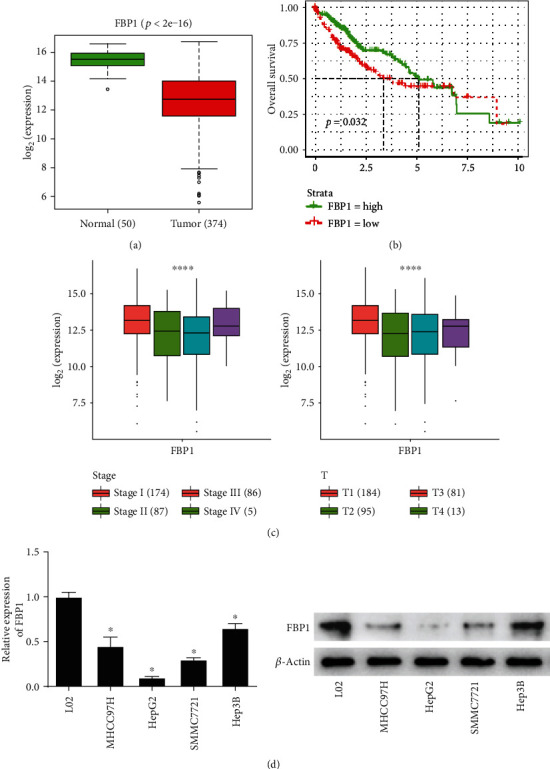
Figure 1.

FBP1 is significantly low in liver cancer cells. (a) Boxplot of the expression of FBP1 in the normal tissue and the tumor tissue samples, with green representing the normal group and red representing the tumor group. (b) Survival curve of FBP1, with abscissa representing the time (unit: year), ordinate representing the survival rate, green curve representing high expression, and red curve representing low expression. (c) Correlation between FBP1 and clinical features. (d) The expression of FBP1 in human normal liver cell line (L02) and 4 human liver cancer cell lines (MHCC97H, HepG2, SMMC7721, and Hep3B); ∗ represents p < 0.05 and ∗∗∗∗ represents p < 0.0001.
