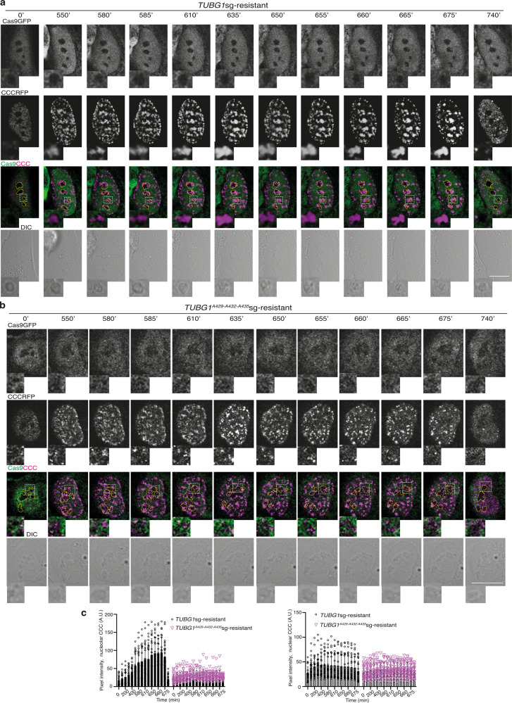
Fig. 7. The γ-tubulin PIP mutant reduced the size of CCC complexes formed in the nucleus.
a, b The DIC/fluorescence images show time-lapse series of a stable TUBGsg-U2OS cell co-expressing either γ-tubulinresist (a) or a γ-tubulinA429-A432-A435resist (b) and cell cycle chromobody (CCCRFP). The image series present chosen frames illustrating the accumulation of CCCRFP and how mutations in the γ-tubulin PIP motif affect the formation of CCCRFP foci (N = 30 cells). Scale bars: 10 μm (see also Supplementary Fig. 9). c The graphs show the time-dependent changes in fluorescence intensity in the nucleolus and nuclear compartment as indicated by the cells in a, b (AU; mean ± SD; N = 22 cells, ***P < 0.001). Source data are provided in Supplementary Data 1.

