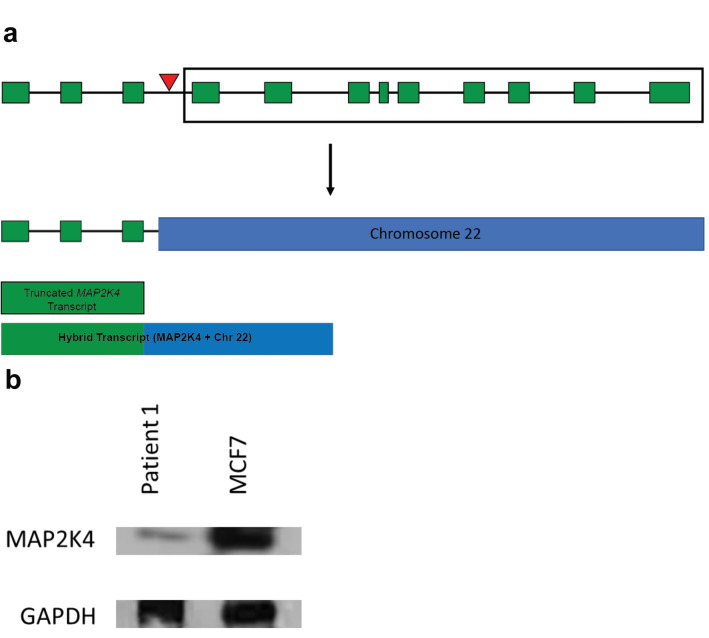
Figure 4.
Translocation within MAP2K4 gene decreases protein production in Patient 1. (A) Schematic of the primary transcript of MAP2K4. The exons of the transcript are shown as green boxes and the red triangle above MAP2K4 depicts the approximate location of the translocation. The translocation results in a truncation of MAP2K4, leaving only the first three exons remaining on Chromosome 17. The portion of MAP2K4 shown in the box is translocated to Chromosome 22. The translocation is depicted below the arrow. Truncated and hybrid transcripts are shown below the translocation. (B) Western blot of MAP2K4 in control MCF7 cells and ascites-derived cells from Patient 1. The control cells show clear expression of MAP2K4, while the cells taken from Patient 1 show decreased production of the protein. GAPDH blotting was performed as a loading control. Uncropped images of the Western blot are shown in Supplemental Figure 7.

