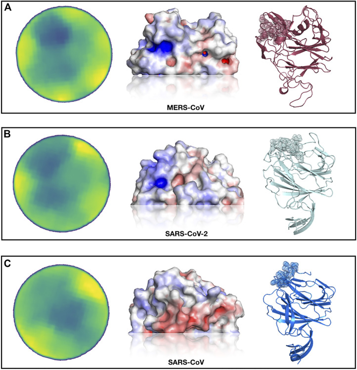
FIGURE 3.
Identification of a SARS-CoV-2 spike region very similar to the sialic-acid binding site on MERS-CoV spike. (A) From left to right, projected region of the real sialic-acid binding site on MERS-CoV, electrostatic potential surface of the same region and cartoon representation of the MERS-CoV spike protein with the binding site highlighted. (B) Putative sialic-acid binding region on SARS-CoV-2 as predicted by our Zernike-based method. From left to right, the projected region of putative interaction site between SARS-CoV and sialic acid, electrostatic potential surface, and cartoon representation of the SARS-CoV spike protein with the binding site highlighted. (C) Same as (B) but for SARS-CoV spike protein.

