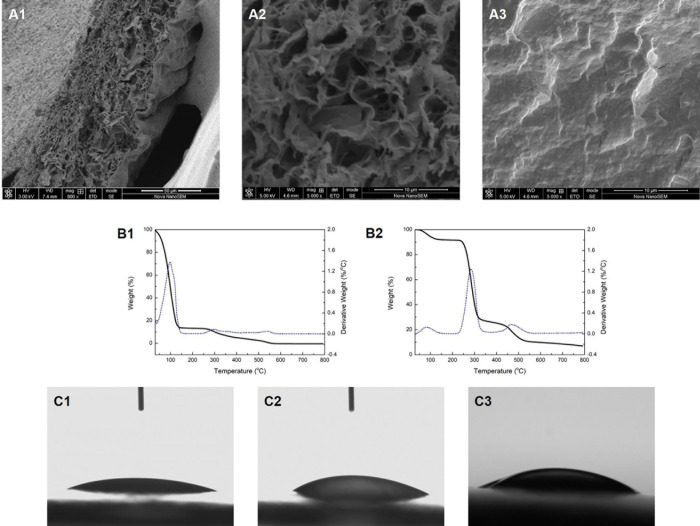
FIGURE 1.
The cross-section morphology of the Col/PVA dual-layer membrane showing (A1) both layers, (A2) collagen layer, (A3) PVA layer. TG analysis of (B1) Col/PVA dual-layer membrane, (B2) pure PVA membrane. WCA of (C1) collagen side, (C2) PVA side of the Col/PVA dual-layer membrane, (C3) pure PVA membrane.

