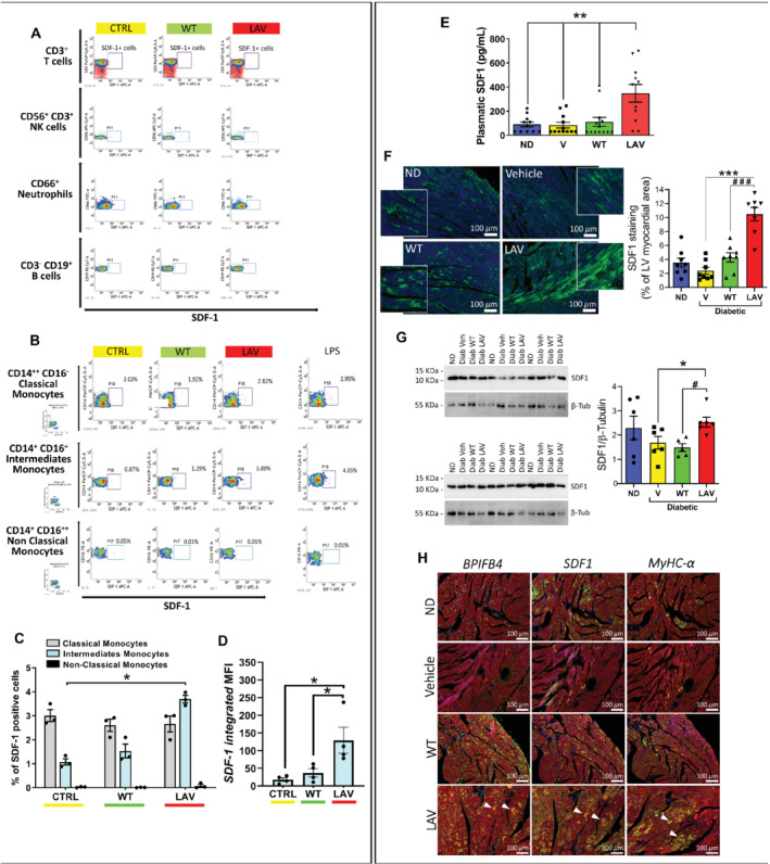
Figure 4.

LAV‐BPIFB4 induces stromal cell‐derived factor‐1 (SDF‐1) in human monocytes in vitro and in peripheral blood and hearts of diabetic mice in vivo. (A–D) Cytofluorimetric analysis of the peripheral blood mononuclear cells (PBMCs) from type 2 diabetic patients incubated with WT‐BPIFB4 or LAV‐BPIFB4 protein or vehicle for 48 h. LPS was used as a positive control stimulation. At the end of the cell culture treatment, PBMCs were recovered, stained for the different antigens and anti‐intracellular SDF‐1, and analysed by flow cytometry. (A) T cells, NK cells, neutrophils, and B cells. (B) Monocytes. (C) Frequency of SDF‐1 positive events within subclasses of monocytes. (D) Relative fluorescence intensity of SDF‐1 in the intermediate monocyte population. Individual values and mean ± SEM. n = 4 biological replicates. *P < 0.05. (E) Immunoreactive SDF‐1 levels in murine peripheral blood. Bar graphs show individual values and mean ± SEM. n = 11–13 mice per group. **P < 0.001 between longevity‐associated variant (LAV) and other groups. (F) Immunohistochemistry (SDF‐1 in green, DAPI in blue, scale bars: 100 μm) and (G) western blot analyses of SDF‐1 expression in the mice heart. Bar graphs show individual values and mean ± SEM. n = 5–8 mice per group. *P < 0.05, ***P < 0.001 vs. vehicle; # P < 0.05, ### P < 0.001 vs. WT. (H) Immunohistochemistry (scale bars: 100 μm) of BPIFB4, SDF‐1 and myosin heavy chain isoform alpha (MyHC‐α) expression (all in green), α‐sarcomeric actin in red, and DAPI blue. Triangles indicate cardiomyocytes co‐expressing the three proteins.
